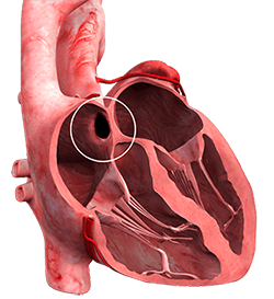
What is an Atrial Septal Defect (ASD)?
An atrial septal defect is a hole in between the left and right upper chambers of the heart. It is a congenital defect, which means it is present at birth. The exact cause of ASD is unknown.
What is an Atrial Septal Defect Closure?
Atrial septal defect closure is the surgical intervention that involves either suturing the edges of the atrial septal defect together or suturing a patch to close up the hole in the heart wall.
What Happens if an Atrial Septal Defect is left Untreated?
Due to the abnormal flow of blood between the right atrium and left atrium, the heart must work harder to pump blood resulting in:
- Enlargement of the right atrium
- Increased pulmonary artery pressure
- Stiffening of the pulmonary artery
- Irregular heart rhythm
- Heart failure
Symptoms of an Atrial Septal Defect
The symptoms may occur at any age of your life. The symptoms include:
- Heart murmur
- Heart palpitations
- Fatigue
- Shortness of breath
- Stroke
- Swelling of hands, legs and abdomen
- A blue tinge of the skin
Diagnosis of an Atrial Septal Defect
Your doctor may suspect an atrial septal defect if a heart murmur is heard on physical examination. One or more of the following tests may also be ordered:
- Echocardiogram
- Electrocardiogram
- An X-ray of your chest region
- Magnetic Resonance Imaging (MRI)
- Computed Tomography (CT) scan
How is Atrial Septal Defect Closure Performed?
Depending on the size of the atrial septal defect, treatment involves cardiac catherisation, minimally invasive surgery, or open-heart surgery. Talk to your surgeon to know which is best for you.
Cardiac Catherisation
It involves the following steps:
- You are given local anaesthesia
- A small incision is made near your groin or thigh region
- A catheter is inserted through a large vein and guided towards your heart
- Your surgeon may insert a mesh or a plug through the catheter to close the hole
- The catheter is removed, and your surgeon closes the incision near your thigh region
Minimal Invasive Surgery
The technique may involve the following steps:
- You are given general anaesthesia
- Your surgeon makes an incision of about 4 cm to 6 cm at the side of your chest
- You are supported with a heart-lung machine for normal respiration and circulation
- A soft retractor is inserted between the ribs
- Your surgeon inserts minimally invasive surgical instruments
- A 3D camera is inserted to view the defect
- Your surgeon inserts a patch to close the defect
- The patch can be a graft taken from the pericardium of your heart or a synthetic material
- The incision is closed with a bandage
Open Heart Surgery
This procedure involves incision of your breastbone to expose the heart. The procedure is rarely performed and is only done in cases where the ASD is very large or in an unusual position.
Postsurgical Recovery after Atrial Septal Defect Closure
During the recovery period, your surgeon will recommend refraining from vigorous physical activity. Once fully healed from the surgery most patients can expect to return to their active lifestyles without any restrictions.
